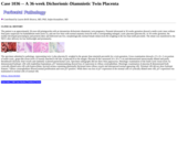
(This case study was added to OER Commons as one of a batch of over 700. It has relevant information which may include medical imagery, lab results, and history where relevant. A link to the final diagnosis can be found at the end of the case study for review. The first paragraph of the case study -- typically, but not always the clinical presentation -- is provided below.)
The patient is an approximately 30-year-old primigravida with an intrauterine dichorionic-diamniotic twin pregnancy. Prenatal ultrasound at 30 weeks gestation showed a multi-cystic mass without fetal parts suspicious for hydatidiform mole (twin A), and one live fetus with normal anatomy (twin B) with a corresponding enlarged, cystic placenta (placenta B). At 36 weeks gestation, the mother developed preeclampsia and preterm labor and delivered one live, morphologically normal female infant (twin B) weighing in the less than tenth percentile. The infant was transferred to the NICU after delivery for low birthweight and prematurity.
- Subject:
- Applied Science
- Education
- Health, Medicine and Nursing
- Life Science
- Material Type:
- Case Study
- Diagram/Illustration
- Provider:
- University of Pittsburgh School of Medicine
- Provider Set:
- Department of Pathology
- Author:
- Lauren Brilli Skvarca
- Stefan Kostadinov
- Date Added:
- 08/01/2022