Updating search results...

Search Resources

739 Results

View
Selected filters:
  • University of Pittsburgh School of Medicine
Pathology Case Study: A male in his 30s with  a mediastinal mass
Read the Fine Print
Some Rights Reserved
Rating
0.0 stars

(This case study was added to OER Commons as one of a batch of over 700. It has relevant information which may include medical imagery, lab results, and history where relevant. A link to the final diagnosis can be found at the end of the case study for review. The first paragraph of the case study -- typically, but not always the clinical presentation -- is provided below.)

A male in his late 30s with a history of chronic alcohol abuse and depression presented the emergency room complaining of severe shortness of breath. A detailed history revealed that he had been relatively healthy until one month prior, when he began to experience progressively worsening shortness of breath. On physical examination, he was diaphoretic and in obvious distress, with a respiratory rate of 36, a heart rate of 120, and a seated blood pressure of 81/32. Decreased breath sounds were noted on the left side of his chest, and his trachea was deviated to the right. He was placed on a 100% non-rebreather mask and an arterial blood gas was taken: pH: 7.07, pCO2: 57, bicarbonate: 15.9, pO2: 96, O2 saturation: 91%. Initial radiographic studies revealed a large left pleural effusion with resultant mediastinal and tracheal shift into the right hemithorax. The patient was intubated and placed on a ventilator. A left chest tube was placed, with immediate drainage of approximately 4 liters of serous fluid. A repeat chest x-ray revealed a large superior mediastinal mass, suspicious for adenopathy or primary mediastinal tumor. He was started on intravenous fluids, antibiotics, and vasopressor support, but his condition continued to worsen. He was transferred to the MICU for further management, but went into cardiac arrest and was pronounced dead upon arrival.

Subject:
Applied Science
Education
Health, Medicine and Nursing
Life Science
Material Type:
Case Study
Diagram/Illustration
Provider:
University of Pittsburgh School of Medicine
Provider Set:
Department of Pathology
Author:
Beth Z Clark
Larry Nichols
Lawrence B Fialkow
Date Added:
08/01/2022
Pathology Case Study: A male in his 40s with  a pancreatic mass
Read the Fine Print
Some Rights Reserved
Rating
0.0 stars

(This case study was added to OER Commons as one of a batch of over 700. It has relevant information which may include medical imagery, lab results, and history where relevant. A link to the final diagnosis can be found at the end of the case study for review. The first paragraph of the case study -- typically, but not always the clinical presentation -- is provided below.)

A male inhis 40s presented to the emergency department with abdominal pain, nausea and vomiting. A computed tomography (CT) scan showed an 8 cm mass in the tail of the pancreas (Fig. 1). He underwent distal pancreatectomy and splenectomy.

Subject:
Applied Science
Education
Health, Medicine and Nursing
Life Science
Material Type:
Case Study
Diagram/Illustration
Provider:
University of Pittsburgh School of Medicine
Provider Set:
Department of Pathology
Author:
Alyssa M Krasinskas
Eizaburo Sasatomi
Date Added:
08/01/2022
Pathology Case Study: A male in his 40s with severely compromised vision
Read the Fine Print
Some Rights Reserved
Rating
0.0 stars

(This case study was added to OER Commons as one of a batch of over 700. It has relevant information which may include medical imagery, lab results, and history where relevant. A link to the final diagnosis can be found at the end of the case study for review. The first paragraph of the case study -- typically, but not always the clinical presentation -- is provided below.)

A male in his 40s with a prior medical history of Behcet's disease and severely compromised vision presented with a cognitive disorder and chorea. His symptoms began 8 years ago, over which time the patient has steadily deteriorated. His mother had similar symptoms and was diagnosed as having Huntington disease. Few family members on the maternal side were also affected (see Pedigree). None of the mother's relatives were previously tested.

Subject:
Applied Science
Education
Health, Medicine and Nursing
Life Science
Material Type:
Case Study
Diagram/Illustration
Provider:
University of Pittsburgh School of Medicine
Provider Set:
Department of Pathology
Author:
Jeffrey Kant
Laurentia Nodit
Melina Flanagan
Date Added:
08/01/2022
Pathology Case Study: A male in his 50s with  chronic anemia
Read the Fine Print
Some Rights Reserved
Rating
0.0 stars

(This case study was added to OER Commons as one of a batch of over 700. It has relevant information which may include medical imagery, lab results, and history where relevant. A link to the final diagnosis can be found at the end of the case study for review. The first paragraph of the case study -- typically, but not always the clinical presentation -- is provided below.)

A caucasian man in his 50s presented to the emergency department with increasing exertional dyspnea, generalized weakness, lethargy, and anemia. The patient underwent orthotopic cardiac transplant approximately six months earlier for ischemic cardiomyopathy secondary to a previous myocardial infarction. The anemia was first noted a few weeks postoperatively.

Subject:
Applied Science
Education
Health, Medicine and Nursing
Life Science
Material Type:
Case Study
Diagram/Illustration
Provider:
University of Pittsburgh School of Medicine
Provider Set:
Department of Pathology
Author:
J Manuel Zarandona
Sandra S Kaplan
Date Added:
08/01/2022
Pathology Case Study: A male in his 60s with plasma cell neoplasm, favor solitary plasmacytoma of bone
Read the Fine Print
Some Rights Reserved
Rating
0.0 stars

(This case study was added to OER Commons as one of a batch of over 700. It has relevant information which may include medical imagery, lab results, and history where relevant. A link to the final diagnosis can be found at the end of the case study for review. The first paragraph of the case study -- typically, but not always the clinical presentation -- is provided below.)

The patient was male in his 60s with a history of monoclonal gammopathy of undetermined significance, who presented with borderline normocytic anemia. Peripheral blood showed hemoglobin 12.4 g/dL, hematocrit 36.3 percent, MCV 86.6 fL, MCH 29.6 pg, and MCHC 34.2 g/dL.

Subject:
Applied Science
Education
Health, Medicine and Nursing
Life Science
Material Type:
Case Study
Diagram/Illustration
Provider:
University of Pittsburgh School of Medicine
Provider Set:
Department of Pathology
Author:
Karen Schodel
Teresa LaCaria
Date Added:
08/01/2022
Pathology Case Study: A male in his 70s with a brain mass
Read the Fine Print
Some Rights Reserved
Rating
0.0 stars

(This case study was added to OER Commons as one of a batch of over 700. It has relevant information which may include medical imagery, lab results, and history where relevant. A link to the final diagnosis can be found at the end of the case study for review. The first paragraph of the case study -- typically, but not always the clinical presentation -- is provided below.)

A male in his 70s with history of Parkinson's disease presented with complaints of double vision and falls on the left side.

Subject:
Applied Science
Education
Health, Medicine and Nursing
Life Science
Material Type:
Case Study
Diagram/Illustration
Provider:
University of Pittsburgh School of Medicine
Provider Set:
Department of Pathology
Author:
Muammar Arida
Date Added:
08/01/2022
Pathology Case Study: A male in his 80s with left sided neck mass
Read the Fine Print
Some Rights Reserved
Rating
0.0 stars

(This case study was added to OER Commons as one of a batch of over 700. It has relevant information which may include medical imagery, lab results, and history where relevant. A link to the final diagnosis can be found at the end of the case study for review. The first paragraph of the case study -- typically, but not always the clinical presentation -- is provided below.)

A male in his late 80s with a past medical history significant for prostatic adenocarcinoma 10 years ago presented with a left sided neck mass. An evaluation by fine needle aspiration was requested. Based on the Diff Quik stain slides (Figures 1, 2 and 3) please answer the following questions:

Subject:
Applied Science
Education
Health, Medicine and Nursing
Life Science
Material Type:
Case Study
Diagram/Illustration
Provider:
University of Pittsburgh School of Medicine
Provider Set:
Department of Pathology
Author:
Fiona Craig
Maurice R. Grant
Date Added:
08/01/2022
Pathology Case Study: A male in his late 60's with recurrent extracerebral tumor
Read the Fine Print
Some Rights Reserved
Rating
0.0 stars

(This case study was added to OER Commons as one of a batch of over 700. It has relevant information which may include medical imagery, lab results, and history where relevant. A link to the final diagnosis can be found at the end of the case study for review. The first paragraph of the case study -- typically, but not always the clinical presentation -- is provided below.)

Over the past year, this man in late sixties had complained about progressive weakness of concentration and memory disturbances, associated with word finding difficulties. Upon admission, neurological examination revealed a distinct paresis of the right arm and leg. Knee and ankle reflexes were brisk on the right side and reduced on the left side. Additionally, anemia and reduced stride length due to known Parkinson´s disease were noted. MRI examination revealed an extra-axial, parasagittal tumor 3cm in diameter located in the left frontoparietal region. The lesion was isointense on T1 and T2-weighted images, with homogeneous signal enhancement after contrast medium application (Figure 1A).

Subject:
Applied Science
Education
Health, Medicine and Nursing
Life Science
Material Type:
Case Study
Diagram/Illustration
Provider:
University of Pittsburgh School of Medicine
Provider Set:
Department of Pathology
Author:
Christian Mawrin
Cordula Scherlach
Elmar Kirches
Knut Dietzmann
Rainer Hahne
Date Added:
08/01/2022
Pathology Case Study: A male teenager with a brain mass
Read the Fine Print
Some Rights Reserved
Rating
0.0 stars

(This case study was added to OER Commons as one of a batch of over 700. It has relevant information which may include medical imagery, lab results, and history where relevant. A link to the final diagnosis can be found at the end of the case study for review. The first paragraph of the case study -- typically, but not always the clinical presentation -- is provided below.)

The patient is a teenager boy who was previously diagnosed with ALL in 2001. He had numerous relapses and received a bone marrow transplant in 2002. He subsequently developed grafts vs host disease and his immunosuppression increased. The patient subsequently presented with visual disturbances and headache over a period of two to three weeks in May 2003. Initially, CT showed a large mass in the in the right temporal lobe. All other radiology was negative. A stereotactic brain biopsy was performed.

Subject:
Applied Science
Education
Health, Medicine and Nursing
Life Science
Material Type:
Case Study
Diagram/Illustration
Provider:
University of Pittsburgh School of Medicine
Provider Set:
Department of Pathology
Author:
Raj Hari
Date Added:
08/01/2022
Pathology Case Study: A man at 70s Had Episodes of Syncopy
Read the Fine Print
Some Rights Reserved
Rating
0.0 stars

(This case study was added to OER Commons as one of a batch of over 700. It has relevant information which may include medical imagery, lab results, and history where relevant. A link to the final diagnosis can be found at the end of the case study for review. The first paragraph of the case study -- typically, but not always the clinical presentation -- is provided below.)

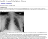
A Caucasian man in his 70's was visiting his neighbors when he had a witnessed syncopal episode. Shortly after, he had a second episode and an ambulance was called. The patient became unresponsive before EMS arrived and required CPR. EMS was able to restore the patient's circulatory function en route to the hospital. Upon arrival in the emergency department, a chest x-ray was obtained.

Subject:
Applied Science
Education
Health, Medicine and Nursing
Life Science
Material Type:
Case Study
Diagram/Illustration
Provider:
University of Pittsburgh School of Medicine
Provider Set:
Department of Pathology
Author:
Jeffrey Nine
Michelle Stram
Date Added:
08/01/2022
Pathology Case Study: A man in his 20s with a sudden onset of polydipsia and polyuria
Read the Fine Print
Some Rights Reserved
Rating
0.0 stars

(This case study was added to OER Commons as one of a batch of over 700. It has relevant information which may include medical imagery, lab results, and history where relevant. A link to the final diagnosis can be found at the end of the case study for review. The first paragraph of the case study -- typically, but not always the clinical presentation -- is provided below.)

The patient is a man in his 20s who experienced a sudden onset of polydipsia and polyuria. Laboratory testing revealed increased serum osmolality (340 mOsm/kg, reference range 275-299), decreased urine osmolality (40 mOsm/kg, reference range 50-1400), and increased serum sodium (150 mEq/L, reference range 136-146). A water deprivation test demonstrated decrease of serum osmolality into normal range with increased urine osmolality in response to a dose of DDAVP, and so the patient was diagnosed with central diabetes insipidus. The initial MRI scan showed mild thickening of the pituitary stalk.

Subject:
Applied Science
Education
Health, Medicine and Nursing
Life Science
Material Type:
Case Study
Diagram/Illustration
Provider:
University of Pittsburgh School of Medicine
Provider Set:
Department of Pathology
Author:
Geoffrey Murdoch
Manuel Zarandona
Date Added:
08/01/2022
Pathology Case Study: A man in his 40s with a 2-year history of epistaxis
Read the Fine Print
Some Rights Reserved
Rating
0.0 stars

(This case study was added to OER Commons as one of a batch of over 700. It has relevant information which may include medical imagery, lab results, and history where relevant. A link to the final diagnosis can be found at the end of the case study for review. The first paragraph of the case study -- typically, but not always the clinical presentation -- is provided below.)

A previously healthy man in his 40s came to clinical attention with a 2-year history of epistaxis. Upon rhinoscopic inspection, an exophytic, red nodule was found in the nasopharynx, centered on the midline. The tumor was covered by intact mucosa and appeared pulsating. Sagittal T1-weighted images (Figure 1) with fat suppression showed a midline mass with strong contrast enhancement, involving the nasopharyngeal space (arrow) and eroding the clivus (arrowhead). Following an incisional biopsy, surgical excision of the tumor was carried out by transfacial approach.

Subject:
Applied Science
Education
Health, Medicine and Nursing
Life Science
Material Type:
Case Study
Diagram/Illustration
Provider:
University of Pittsburgh School of Medicine
Provider Set:
Department of Pathology
Author:
Ildiko Szalay
Jacqueline Delavelle
Pier Luigi Di Patre
Date Added:
08/01/2022
Pathology Case Study: A man in his 40s with headaches and dyspnea
Read the Fine Print
Some Rights Reserved
Rating
0.0 stars

(This case study was added to OER Commons as one of a batch of over 700. It has relevant information which may include medical imagery, lab results, and history where relevant. A link to the final diagnosis can be found at the end of the case study for review. The first paragraph of the case study -- typically, but not always the clinical presentation -- is provided below.)

A 40 year old man presented with severe frontal headaches, dry cough, impaired balance and dizziness for six weeks. On examination he had low-grade pyrexia and investigations revealed mild lymphopenia. Brain CT was normal and chest x-ray showed right hilar adenopathy with bilateral parenchymal infiltrates. A diagnosis of atypical pneumonia was made.

Subject:
Applied Science
Education
Health, Medicine and Nursing
Life Science
Material Type:
Case Study
Diagram/Illustration
Provider:
University of Pittsburgh School of Medicine
Provider Set:
Department of Pathology
Author:
Browne LI
Delanty N
Lane H
O Neill S
Date Added:
08/01/2022
Pathology Case Study: A man in his 40s with severe headaches after a fall
Read the Fine Print
Some Rights Reserved
Rating
0.0 stars

(This case study was added to OER Commons as one of a batch of over 700. It has relevant information which may include medical imagery, lab results, and history where relevant. A link to the final diagnosis can be found at the end of the case study for review. The first paragraph of the case study -- typically, but not always the clinical presentation -- is provided below.)

The patient was a male in his 40s with a history of asthma, smoking and possible ethanol abuse, who was otherwise in good health and not on any medication. The patient suffered a traumatic injury to his head after falling down a flight of stairs, while under the influence of alcohol, on the evening of Day 1. It is unclear whether or not the patient suffered a loss of consciousness at that time; he went to bed and complained of a severe headache when awakened around noon the next day

Subject:
Applied Science
Education
Health, Medicine and Nursing
Life Science
Material Type:
Case Study
Diagram/Illustration
Provider:
University of Pittsburgh School of Medicine
Provider Set:
Department of Pathology
Author:
& William Pasculle
Larry Nichols
Marta Couce
ScD
Suzanne Bakdash
Date Added:
08/01/2022
Pathology Case Study: A man in his 50s with  fever, headache, and sore throat
Read the Fine Print
Some Rights Reserved
Rating
0.0 stars

(This case study was added to OER Commons as one of a batch of over 700. It has relevant information which may include medical imagery, lab results, and history where relevant. A link to the final diagnosis can be found at the end of the case study for review. The first paragraph of the case study -- typically, but not always the clinical presentation -- is provided below.)

The patient is a Caucasian male in his 50s with a past medical history of hypertension complaining of "fever, headache, and sore throat." The patient stated that his symptoms began with a sore throat approximately five days prior to admission, followed by fever with a Tmax of 38oC and headache one day prior to admission. He described the headache as moderate to severe in intensity, sharp in nature, and unilateral, being present on the right maxillary and mandibular areas of his face, with radiation to the right neck. He denied cough, dyspnea, photophobia, rash, rhinorrhea, dysphagia, diarrhea, recent travel or sick contacts.

The patient's only outpatient medications are anti-hypertensives. He has chewed tobacco for over 30 years. He denied intravenous or other illicit drug use. Family history is noncontributory.

Physical examination revealed a temperature of 38.3oC, respiratory rate 15, pulse 99, blood pressure 182/110 and pulse oximetry 98% on room air. The remainder of the exam was negative except for poor dentition and pain with palpation of the right mandible and right submandibular region. There was no meningismus, neurologic deficits, or heart murmurs.

Laboratory results were significant for a white blood cell count of 12.5 x 10⁹/L with 91% neutrophils and 4% lymphocytes.

Subject:
Applied Science
Education
Health, Medicine and Nursing
Life Science
Material Type:
Case Study
Diagram/Illustration
Provider:
University of Pittsburgh School of Medicine
Provider Set:
Department of Pathology
Author:
Nicole Esposito
Date Added:
08/01/2022
Pathology Case Study: A man in his 50s with progressive aphasia and right-sided hemiplegia
Read the Fine Print
Some Rights Reserved
Rating
0.0 stars

(This case study was added to OER Commons as one of a batch of over 700. It has relevant information which may include medical imagery, lab results, and history where relevant. A link to the final diagnosis can be found at the end of the case study for review. The first paragraph of the case study -- typically, but not always the clinical presentation -- is provided below.)

A man in his 50s was diagnosed with Glioblastoma Mulitforme of the left temporal lobe for which he underwent chemotherapy and external beam radiation therapy. He presented suddenly nine months later with progressive aphasia and right-sided hemiplegia. Following a physical and neurological examination it was noted that the patient again had a mass situated in the left temporal lobe (in the previous site of the surgery) associated with significant compression of the brainstem.

Subject:
Applied Science
Education
Health, Medicine and Nursing
Life Science
Material Type:
Case Study
Diagram/Illustration
Provider:
University of Pittsburgh School of Medicine
Provider Set:
Department of Pathology
Author:
Leena Lourduraj
Date Added:
08/01/2022
Pathology Case Study: A man in his 70's who has monoclonal gammopathy of undetermined significance
Read the Fine Print
Some Rights Reserved
Rating
0.0 stars

(This case study was added to OER Commons as one of a batch of over 700. It has relevant information which may include medical imagery, lab results, and history where relevant. A link to the final diagnosis can be found at the end of the case study for review. The first paragraph of the case study -- typically, but not always the clinical presentation -- is provided below.)

The patient was a man in his 70s with a history of right lower extremity deep venous thrombosis with multiple pulmonary emboli with right lower lobe pulmonary infarction (most recent hospitalization), asthma, benign prostatic hypertrophy, and intermittent vertigo.

Subject:
Applied Science
Education
Health, Medicine and Nursing
Life Science
Material Type:
Case Study
Diagram/Illustration
Provider:
University of Pittsburgh School of Medicine
Provider Set:
Department of Pathology
Author:
Beth Clark
Bruce Rabin
Date Added:
08/01/2022
Pathology Case Study: A man in his 70s with nausea and vomiting
Read the Fine Print
Some Rights Reserved
Rating
0.0 stars

(This case study was added to OER Commons as one of a batch of over 700. It has relevant information which may include medical imagery, lab results, and history where relevant. A link to the final diagnosis can be found at the end of the case study for review. The first paragraph of the case study -- typically, but not always the clinical presentation -- is provided below.)

This elderly man had a medical and oncologic history significant for a myeloproliferative disorder in the remote past, chronic lymphocytic leukemia, recurrent osteochondroma, prostatic adenocarcinoma, renal insufficiency and coronary artery disease. Past surgical history is notable for a left nephrectomy for "benign" renal disease 28 years earlier and partial colectomy for severe polyposis. He presented for total hip replacement due to worsening hip pain unresponsive to analgesic treatments leaving him virtually bedridden. Several days before his admission, he developed nausea and vomiting with loss of appetite and indeterminate weight loss. Physical examination showed only a slightly enlarged prostate and an abdominal hernia. No abdominal masses or lymphadenopathy were appreciated. Laboratory evaluation then included a complete blood count showing a white blood cell count of 50,700/cu mm with a differential of 6% polymorphonuclear leucocytes, 89% lymphocytes, 3% monocytes, 1% eosinophils, and 1% basophils. Hemoglobin was 11.6 g/dl and platelet count 168,000/cu mm. Electrolytes were within normal limits. Blood urea nitrogen was 32 mg/dl and creatinine 1.7 mg/dl. Liver enzymes, alkaline phosphatase, and total bilirubin were within normal limits. Albumin was marginally low at 3.1 g/dl. Prostatic specific antigen level was 0.1 ng/ml. An abdominal and retroperitoneal ultrasound was performed revealing a 12 cm by 9.5 cm lobulated, hypoechoic soft tissue mass with central necrosis in the right suprarenal region. Subsequent computed tomography scan demonstrated a 10.3 cm by 9.3 cm lobulated heterogeneous right adrenal mass. The mass displaced the right kidney posteriorly and inferiorly with focal compression of the inferior vena cava and extending to the renal hilum (Figures 1A, 1B, and 1C). CT guided biopsy of the mass was diagnosed as cytologically and histologically bland adrenal cortical tissue. The patient subsequently underwent exploratory laparotomy with resection of the adrenal mass. Intraoperatively, a large necrotic appearing tumor encompassed the right adrenal gland, but extended to and was fixed to the inferior vena cava. The tumor was also seen to extend under the right renal vein and involve the right renal hilum. The tumor was incompletely resected secondary to fixation to adjacent structures and intraoperative hemorrhage. This final pathologic diagnosis in this case was based on a compilation of pathologic data including immunohistochemistry, electron microscopy, and mutational profiling.

Subject:
Applied Science
Education
Health, Medicine and Nursing
Life Science
Material Type:
Case Study
Diagram/Illustration
Provider:
University of Pittsburgh School of Medicine
Provider Set:
Department of Pathology
Author:
Eizaburo Sasatomi
John A Ozolek
Patricia A Swalsky
Rajiv Dhir
Sydney D Finkelstein
Date Added:
08/01/2022
Pathology Case Study: A man in his 70s with right upper quadrant and left-sided back pain
Read the Fine Print
Some Rights Reserved
Rating
0.0 stars

(This case study was added to OER Commons as one of a batch of over 700. It has relevant information which may include medical imagery, lab results, and history where relevant. A link to the final diagnosis can be found at the end of the case study for review. The first paragraph of the case study -- typically, but not always the clinical presentation -- is provided below.)

A man in his 70s presented with right upper quadrant and left-sided back pain. He underwent a series of evaluations including a CT scan of the abdomen and pelvis, which revealed an incidental soft tissue density in the tail of the pancreas with likely involvement of the splenic hilum tracking along the splenic artery. A subsequent MRI confirmed the soft tissue density in the distal pancreas along with possible splenic vein thrombosis. Distal pancreatectomy, splenectomy, and partial omentectomy were performed and the specimen was sent for histopathologic evaluation.

Subject:
Applied Science
Education
Health, Medicine and Nursing
Life Science
Material Type:
Case Study
Diagram/Illustration
Provider:
University of Pittsburgh School of Medicine
Provider Set:
Department of Pathology
Author:
Kenneth Clark
Sourav Ray
Date Added:
08/01/2022
Pathology Case Study: A man in his early 20s with a mass on the lateral aspect of his right lower leg
Read the Fine Print
Some Rights Reserved
Rating
0.0 stars

(This case study was added to OER Commons as one of a batch of over 700. It has relevant information which may include medical imagery, lab results, and history where relevant. A link to the final diagnosis can be found at the end of the case study for review. The first paragraph of the case study -- typically, but not always the clinical presentation -- is provided below.)

A young man in his early 20s presented to an orthopedic oncologist for evaluation of a mass on the lateral aspect of his right lower leg. He gave a history of the mass growing slowly but progressively over the course of approximately one year. The lesion itself was non-tender, but did cause the patient some foot and ankle pain when he stood for long periods of time, and caused intermittent tingling of the leg and foot distally. The patient denied any history of trauma or prior surgical intervention at the site.

Subject:
Applied Science
Education
Health, Medicine and Nursing
Life Science
Material Type:
Case Study
Diagram/Illustration
Provider:
University of Pittsburgh School of Medicine
Provider Set:
Department of Pathology
Author:
Juan Xing
Thomas Pearce
Date Added:
08/01/2022