
Worksheet that goes over the types of transport associated with a cell membrane.
- Subject:
- Applied Science
- Biology
- Life Science
- Material Type:
- Homework/Assignment
- Author:
- Lisa Zung
- Date Added:
- 07/02/2020

Worksheet that goes over the types of transport associated with a cell membrane.

This is a video and worksheet covering the basics of cell membranes (Anatomy and Physiology 2e, Chapter 3).

This course explores the major areas of cellular and molecular neurobiology, including excitable cells and membranes, ion channels and receptors, synaptic transmission, cell-type determination, axon guidance, neuronal cell biology, neurotrophin signaling and cell survival, synapse formation and neural plasticity. Material includes lectures and exams, and involves presentation and discussion of primary literature. It focuses on major concepts and recent advances in experimental neuroscience.

Outlines the proposed structure and properties of the plasma membrane in mammalian cells and identifies cell compartments that are separated from each other by at least one plasma membrane.
Andy Meal – lecturer in Biological Sciences, School of Nursing, Midwifery and Physiotherapy, University of Nottingham

As taught Semesters 1 and 2, 2011
This learning object describes the structure and function of mammalian cell organelles. It is used as part of the level 1 Biological Sciences module delivered by the School of Nursing, Midwifery and Physiotherapy.
Dr Andy Meal, Lecturer in Biological Sciences, School of Nursing, Midwifery and Physiotherapy

Cells Grade Level: 10th-12thSubject: ALS AnimalsDuration: 100 minutesDOK Level: 3SAMR Level: RedefinitionIndiana Standard: ALSA-2.10 Compare and contrast animal, plant, and bacterial cells at the biological and chemical levels.Objective: Students will be able to compare and contrast animal, plant, and bacterial cells with 95% accuracy.Essential Question: What are the shared components of cellsProcedure: Ask the students what is the basis of all life?Show them the video Cells Cells - Parts of the Cell RapHand out the plant and animal cell worksheets through google classroomHave the students use the following animation CELLS alive! Interactive Animal and Plant Cells to fill out the worksheetsGroup student into pairsExplain that they will be creating a model that compares plant and animal cellsHandout the Cell Model Sheet through google classroomHave the students create a brainstorming sheetProvide one class period for the students to build and fill out the cell model sheet based on their modelsHave the students complete the memory activity Product or Assessment: The students will be assessed on the cell model sheet Credits:Cells Alive Website, http://www.cellsalive.com/cells/cell_model.htm
This is an inquiry activity that uses either pictures of cells or microscopes and slides to get students thinking about cell structure and function. You could look at prokaryotic vs eukaryotic cells, plant vs animal or different types of cells found in one organism. The objective is to have students begin to make the connection between sructure and function in cells.
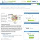
In this lesson, students learn about the basics of cellular respiration. They also learn about the application of cellular respiration to engineering and bioremediation. And, students are introduced to the process of bioremediation and several examples of how bioremediation is used during the cleanup of environmental contaminants.
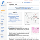
It is important that all aspects of haemostasis can be independently evaluated. This will help to identify the phase affected and to pinpoint what the abnormality is. There are tests available to assess primary haemostasis, secondary haemostasis and fibrinolysis.
The lesson is designed for fourth grade students using scientific investigation to identify and compare the basic parts of a plant and animal cells. This activity consists of several steps over a period of several days. It includes student use of microscopes for observations and journals to collect data to identify and classify the basic parts of plant and animal cells. The assessment has an individual component and uses student work.

Host Harry Kreisler Welcomes Professor Sir John Gurdon for a discussion of advances in research on cell biology. Sir John reflects on his career as a scientist including his path breaking research on cloning. He offers insights into the implications of the revolution in the biological sciences.(46 min)

Host Harry Kreisler welcomes biophysicist Chikashi Toyoshima for a discussion of his remarkable achievement in capturing the first images of cellular activity as the calcium ion pump makes possible the contraction and relaxation of muscle tissue on a signal from the brain. Topics covered include: how structure determines process in cellular activity, the technical innovations required for this breakthrough in the biosciences, and why visualization is so important in understanding cellular processes. He also reflects on creativity in science and how the images he captured affected his understanding of humankind's place in the universe. (52 minutes)

In this visualization adapted from the University of Massachusetts Medical School, discover the role that dengue viral proteins play in a human cell as the virus prepares to replicate.

As taught Semesters 1 and 2, 2011
This learning object describes As taught Semesters 1 and 2, 2011
The learning object distinguishes between some examples of different types of cells on the basis of their microscopic appearance. The learning object is used as part of the level 1 Biological Sciences module delivered by the School of Nursing, Midwifery and Physiotherapy.
Dr Andy Meal, Lecturer in Biological Sciences, School of Nursing, Midwifery and Physiotherapy

Students make edible models of algal cells as a way to tangibly understand the parts of algae that are used to make biofuels. The molecular gastronomy techniques used in this activity blend chemistry, biology and food for a memorable student experience. The models use sodium alginate, which forms a gel matrix when in contact with calcium or moderate acid, to represent the complex-carbohydrate-composed cell walls of algae. Cell walls protect the algal cell contents and can be used to make biofuels, although they are more difficult to use than the starch and oils that accumulate in algal cells. The liquid juice interior of the algal models represents the starch and oils of algae, which are easily converted into biofuels.
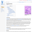
Erythrocytes contain no nucleus and are thus only produced from stem cells. During the fetal stage production is in both the liver and spleen however production is transferred to the bone marrow (red marrow) in the final stages of gestation. Initially erythropoiesis occurs in all bones, however after puberty production is limited to membranous bones (ribs, vertebrae, pelvic bones etc.) as the long bones contain adipose tissue in place of red marrow.
This unit plan was developed by Ochre Education and science teacher Darcie Clarke. It outlines her approach to teaching a Year 8 unit of science on cells, organs and tissues. It includes guidance on how the unit was structured and sequenced and can be used while interacting with the Ochre resources. The plan also allows teachers to see an example of planning for a sequence of lessons and reflect on their own teaching and effective practice. The unit plan is annotated to explicitly show some of the decisions that are made during the planning process.

The fluid mosaic model describes the cell membrane as a tapestry of several types of molecules (phospholipids, cholesterols, and proteins) that are constantly moving. This movement helps the cell membrane maintain its role as a barrier between the inside and outside of the cell environments.

In this video segment adapted from NOVA scienceNOW, hydrogen fuel cell cars promise pollution-free driving, but will we see them anytime soon?
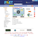
Express yourself through your genes! See if you can generate and collect three types of protein, then move on to explore the factors that affect protein synthesis in a cell.