
Understanding the structure of a skeletal muscle cell.. Created by Raja Narayan.
- Subject:
- Anatomy/Physiology
- Biology
- Life Science
- Material Type:
- Lesson
- Provider:
- Khan Academy
- Provider Set:
- Khan Academy
- Author:
- Raja Narayan
- Date Added:
- 09/18/2013

Understanding the structure of a skeletal muscle cell.. Created by Raja Narayan.
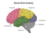
Basic anatomy of the brain: cross section of brain.
A lesson plan for Grade 7 English Language Development and Science. Students develop their knowledge of the circulatory system by studying the structure and function of the heart and its vessels.

Hello Readers, My name is Eddie Joo and I am the co-author as well as the person behind the cover art for this OER. The reason I chose the specific elements on the cover are relatively simple. I dislike the textbooks that just slap random images or things related to the subject and make the cover look really busy. I don't know about you, but any poster or ad that has something to do with Alaska usually includes mountains so I chose the ocean/water instead. Same reason with the animals as the Bear, Wolf, etc. are the go to animals when portraying Alaska so a Dall Sheep, Orca, and an Owl grace the cover. Last but most important to me is the sunrise/sunset gradient. I love seeing both in the winter and is special to me so I added it in as a little Easter egg for me. I hope you enjoy this book! Regards, Eddie
Long Description:
This book is the first edition (Fall 2022) to the exploration of two main core concepts in physiology; Systems Integration, and Homeostasis, in animals at the tissue and organ system level. The flow of this book focuses on how organ systems work together. Foundational homeostatic principles are introduced within the first chapter and threaded throughout the chapters within this text. This text was created by Eddie Joo to supplement students learning within the University of Alaska Anchorage’s Animal Physiology course. The content in this open textbook was adapted from other open textbooks (CC-BY-4.0) resources or created/written by Rachael Hannah and Eddie Joo. This work was funded by an Institutional Development Award (IDeA) from the National Institutes of Health as a Curriculum grant from ALASKA INBRE.
Word Count: 53586
(Note: This resource's metadata has been created automatically by reformatting and/or combining the information that the author initially provided as part of a bulk import process.)
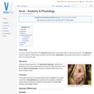
The anus is the terminal portion of the alimentary tract which communicates with the external environment. Two sphincters control it's aperture. It allows faeces and gas to leave the body. Defeacation is the process where faeces are expelled from the rectum through the anus.
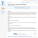
After emerging from the heart, the aortic artery divides into the right and left dorsal branches. Each branch feeds into a set of arches which are unique to the embryo. Most higher vertebrates have have 6 pairs of aortic arches. In the mammal the 5th pair do not form. These arches evolve to form some of the structures of the mammalian circulation. The fate of each arch varies.
The structure of the human body is usually considered in courses in gross anatomy. These courses typically consist of lectures by the faculty, readings in assigned textbooks and the study of photographs and illustrations in human anatomy atlases. However, as student’s progress through the curriculum and move into the more clinical or practice oriented phases, many discover that the anatomical knowledge they actually need is somewhat different from the kind they possess. What many encounter is difficulty in applying their knowledge to a clinical setting.
Applied Human Anatomy was created to better integrate material that is more often than not treated separately in contemporary health care curricula. It is hoped that through this integration students will develop a deeper and more lasting knowledge and understanding of human anatomy as they are likely to need it in the evaluation and management of patients.
A print version is available for purchase on Amazon.
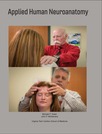
This 155-page manual is comprised of two types of learning activities: 1) free response fill-in-the blank questions focused of the facts and principles of neuroanatomy and neurophysiology that underpin the neurologic examination and specifically developed exercises that demonstrate how the facts and principles are related to the particular tests and procedures that comprise the neurologic examination. Free response questions form the bulk of the Neuroscience Review section of each chapter and are intended as a review of information previously or concurrently being learned regarding the structure, function and organization of the nervous system. Some questions focus on anatomical or physiological facts and relationships that help explain why certain techniques are performed as they are, such as why non-nociceptive tactile stimuli are required in order to activate nerve impulse transmission in the lemniscal system. Other questions are intended to revisit facts and concepts that are needed to properly interpret the elicited findings. 2) The Application Exercises of each chapter are designed to demonstrate how neuroanatomical and neurophysiological information is used in the design of particular clinical tests of neurologic function. The application exercises are also intended to help users learn how to perform and become comfortable with the various clinical maneuvers and tests that comprise the routine neurologic examination. An important outcome of performing these exercises is that, as a member of a learning group, each individual has the opportunity to experience the neurologic examination from the point of view of the subject (patient)—an experience that arguably provides insight and understanding that can be gained in no other way.
Arterial blood gases, basic overview.
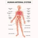
Basic anatomy of the arteries of the human body.
Although the information on this page is based around the anatomy of the canine hindlimb, it is essentially the anatomy of the arteries in domestic species. Any major differences will be discussed on their respective pages

This resource is a video abstract of a research paper created by Research Square on behalf of its authors. It provides a synopsis that's easy to understand, and can be used to introduce the topics it covers to students, researchers, and the general public. The video's transcript is also provided in full, with a portion provided below for preview:
"Ischemic heart disease is a narrowing of the arteries that supply blood and oxygen to the heart. Caused by a build-up of plaque, this restricted blood flow, or ischemia, can cause chest pain and heart attacks, and is among the leading causes of death worldwide. Increasingly, physicians are looking to use MRI to visualize and measure the extent and severity of ischemia. This technique involves the injection of a contrast agent into a blood vessel in the arm. MRI is then used to create a series of images of the heart as the agent is delivered to the muscle, allowing physicians to visualize regions receiving inadequate blood flow. Images are acquired both at rest and during the “stress” induced by giving the patient a drug that increases blood flow to the heart as would occur during exercise. The utility of this approach, however, has been hindered by a lack of objective evaluation methods and the time-consuming task of processing the results in a laboratory setting..."
The rest of the transcript, along with a link to the research itself, is available on the resource itself.
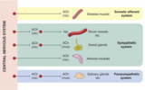
Basic overview of the Autonomic Nervous system along with their respective receptors and neurotransmitters.
The peripheral nervous system found in most domestic species can be segregated into three sub-systems; the sensory system, the somatic motor system and the autonomic system. The autonomic nervous system (ANS) regulates the internal environment of the body including factors such as body temperature, blood pressure and concentrations of many substances. The ANS is also responsible for mobilising the body's resources during stressful situations. The ANS controls gland cells, cardiac muscle cells and smooth muscle cells. Control of this nervous system is involuntary and regulation is via autonomic reflexes. The autonomic reflex arc system is very similar to that of the somatic motor system, i.e. there are sensory (afferent) nerve fibres, an information integration centre, motor (efferent) fibres and effector cells. Any levels of increased activity within the autonomic nervous system can result in both stimulation or inhibition of effector cells, although it is only the efferent part of the reflex arc that is actually considered autonomic.

Hank takes you on a tour of your two-part autonomic nervous system. This episode explains how your sympathetic nervous system and parasympathetic nervous system work together as foils, balancing each other out. Their key anatomical differences - where nerve fibers originate and where their ganglia are located - drive their distinct anatomical functions, making your sympathetic nervous system the "fight or flight" while your parasympathetic nervous system is for "resting and digesting."
Chapters:
Introduction: Autonomic Nervous System
Sympathetic & Parasympathetic Nervous Systems
Origins - Comparing the Sympathetic & Parasympathetic Nervous Systems
Ganglia - Comparing the Sympathetic & Parasympathetic Nervous Systems
Axon Lengths - Comparing the Sympathetic & Parasympathetic Nervous Systems
Review
Credits
Short video on Autonomic nervous system for Anatomy and Physiology.
The intestines occupy the caudal part of the body. They contact the reproductive organs and the gizzard. The small intestine is long and relatively uniform in shape and size. There is no demarcation between the jejunum and the ileum.
The avian respiratory system contains some fundamental differences to the mammalian system.
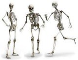
This lesson introduces students to the bones of the axial skeleton, including the bones of the skull, spine, and rib cage. Interactive student and teacher notes are included, along with a Quizlet set, and bookwork assignment to reinforce content.
This is a full Common Course Cartridge shell for BIO 201 Anatomy & Physiology I.Updated July 2023: Updated the labs and added homework from OpenStax Assignable.